05.07.2021 – 18:39
Magnetic resonance imaging (MR) is a painless, non-invasive, and radiation-free medical imaging test used to diagnose small bowel problems. As a specialized form of magnetic resonance imaging (MRI), the test provides detailed images of the intestine through the use of a strong magnetic field.
Purpose of the Test
With MR enterography, the doctor can take high-resolution images of the small intestine to help you diagnose the disease, diagnose and monitor treatment.
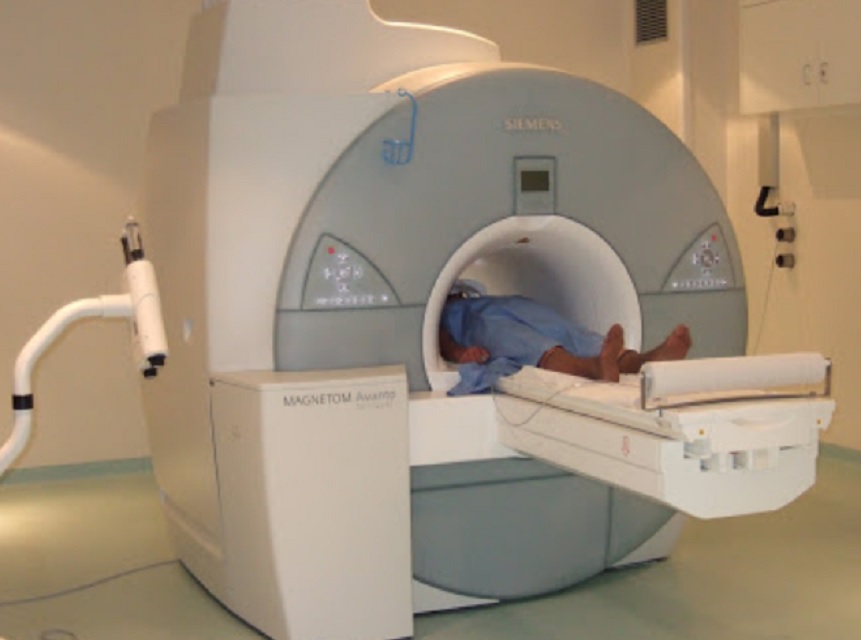
The procedure is done on an MRI machine, which uses powerful magnets to produce a strong magnetic field that helps create detailed computerized images.
MR enterography is performed with a contrast material, which is a fluid that helps to improve image quality. Contrast material is administered orally and / or intravenously.
Since there is no ionizing radiation involved in MR enterography, the procedure can be used – but not preferred – to evaluate young people with inflammatory bowel disease and those with certain types of inflammatory bowel disease. This is because MR enterography can help reduce lifelong exposure to ionizing X-ray radiation.
Diagnosing
Doctors use MR enterography to diagnose a number of medical conditions affecting the small intestine, including inflammatory bowel disease (such as Crohn’s disease).
In addition, MR enterography can identify the following problems:
Inflammation
Internal bleeding
Vascular abnormalities
Tumors
abscesses
Small cracks in the intestinal wall
Small bowel polyps
Intestinal blockages
mONITORING
MR enterography can also be used to find out how well certain treatments are working, and to detect any complications.
Differences and limitations
Unlike a computed tomography (CT) scan (sometimes referred to as computed tomography or CAT scan), MR enterography does not use X-rays to produce images.
Moreover, the contrast material used in MR enterography is usually considered less likely to produce an allergic reaction than the iodine-based contrast materials used for conventional X-rays and CT scan.
In many cases, MR enterography provides a clearer differentiation between abnormal and normal tissue (compared to conventional X-rays and CT scan).
However, performing MR enterography takes much longer than CT enterography (30 to 45 minutes, compared to two to four minutes).
One of the limitations of MR enterography is that patient movement can affect the quality of the images produced. This means that high quality images are only achieved when the person remains completely still and adheres to the breathing instructions during the image recording process. Because people with anxiety may find it difficult to hold their breath, it is often recommended that such people take a sedative before undergoing MR enterography.
Another limitation of MR enterography is that particularly elderly individuals may not fit into the opening of some MRI machines.
Risks and Contraindications
Although MR enterography does not use ionizing radiation, it does use a strong magnetic field. For this reason, it is important to inform your doctor if you have any equipment, implants or metal in your body, or if you have worked with metal in the past. People with certain implants cannot do this procedure, so be sure to notify doctors before an MR registration to make sure it is safe for you.
Magnetic fields can cause some medical devices to malfunction.
Here are some other things to consider before switching to MR enterography:
It is important to tell your radiologist if you have a history of kidney disease, have other health problems, or if you have recently had surgery or medical treatment.
There is a very small risk of an allergic reaction when injecting contrast material. These reactions are usually mild and are relieved with medication. Tell your doctor if you notice any allergic symptoms.
While MR enterography is not known to harm fetuses, it is recommended that pregnant women avoid having any type of MRI exam as a precaution, especially during the first trimester (unless medically necessary).
Patients with very poor renal function and those seeking dialysis face the risk of a rare complication called systemic nephrogenic fibrosis due to the contrast material. If you have a history of kidney disease, you will need to undergo a test to assess if your kidneys are functioning properly.
Some people should not undergo MR enterography. These include individuals with:
Cochlear (ear) implants
Certain types of clips used for brain aneurysms
Certain types of metal wrappers placed inside blood vessels
Almost all cardiac defibrillators and pacemakers.
Some people who have worked with metal in the past may not be able to undergo MR enterography.


